A. Nanotheranostics of infectious diseases
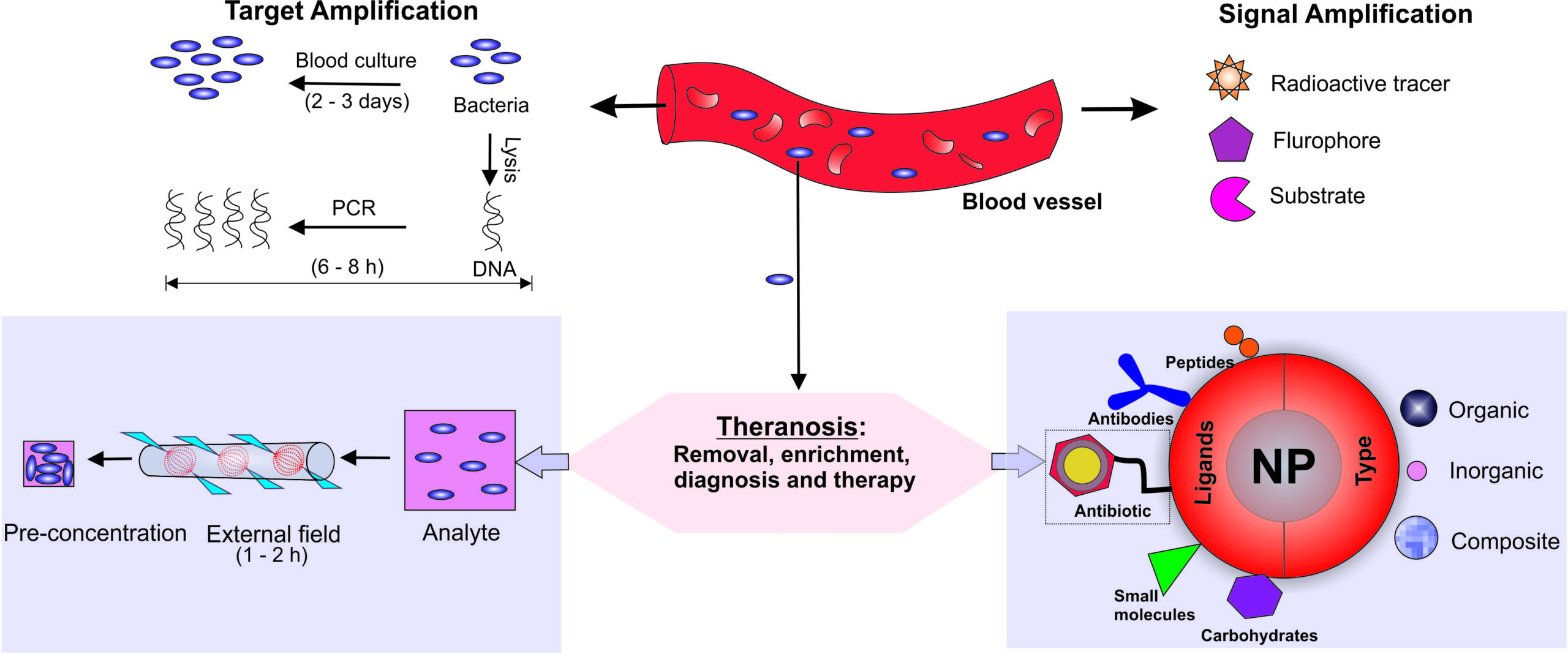
Bloodstream bacterial infections are a serious threat to global public health and economy. The recent figures released by National Center for Health Statistics (NCHS) in 2011 indicate that more than a million people in the US alone get affected by it each year and the sepsis mortality rate is about 28 to 50%. Robust and affordable point-of-care (POC) medical technologies are, therefore, urgently needed for rapid decision-making to initiate proper line of treatment. Current techniques based on blood culture and serology do not have quick turnaround times or adequate sensitivities for early intervention. Moreover, antimicrobial resistance (AMR) poses a great challenge in the fight towards effective bacterial infection management. Nanotheranostics is emerging as a novel strategy combining solutions for rapid diagnosis and treatment in a more personalized way and a large part of our group’s efforts are directed toward this endeavor.
Overview of the nanotheranostic approaches taken by our group
- Prasad P., Sachan S., Suman S., Swyambhu G. and Gupta S., ‘Regenerative core-shell nanoparticles probes for simultaneous removal and detection of endotoxins‘, Langmuir, 34, 7396–7403 (2018)
- Jagtap P., Venkatraman S. and Gupta S., ‘Nanotheranostic Approaches for the Management of Bloodstream Bacterial Infections’ Nanomedicine: Nanotchnology, Biology, and Medicine, 13, 329 – 341 (2016)
Project 1: Endotoxin assay for early bedside diagnosis of septicemia
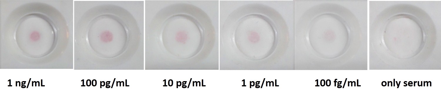
Acute bacterial infections alter the intestinal permeability resulting in the release of intestinal bacteria and their cell wall components like lipopolysachharide (LPS) or lipotechoic acid (LTA) into the bloodstream. This triggers a systemic inflammatory response in the body complicated by organ dysfunction known as sepsis. Sepsis is currently diagnosed based on clinical symptoms and non-specific immunological parameters. A more accepted surrogate biomarker that displays greater sensitivity toward classifying patients with sepsis is procalcitonin (PCT) but its each test is prohibitively expensive. We believe that since LPS and LTA are pathogen-associate molecular patterns (PAMPs), they can serve as important biomarkers for rapid and reliable detection for diagnosis of sepsis especially at early stages. Therefore, in collaboration with Dr. Venkataraman Sritharan at Global Hospitals Hyderabad, we are working on developing an affordable disposable assay that can be used for stratifying Gram-status of bacterial infections within 5 min from a drop of critically-ill patient’s blood, right by their beside. This approach involves working with paper microfuidics, nanoparticle synthesis, novel bioconjucation chemistries and identifying new drug/ligand targets for in vitro diagnostics.
Septifo assay: Gradient colorimetric spots obtained with varying LPS concentrations in human serum.
- Manoharan H., Kalita P., Gupta S. and Sai V.V.R., ‘Plasmonic biosensors for bacterial endotoxin detection on biomimetic C-18 supported fiber optic probes‘, Biosensors and Bioelectronics, 129, 79-86 (2018)
- Jagtap P.*, Singh R.*, Deepika K., Sritharan V., Gupta S., ‘A flow through assay for rapid, bedside stratification of endotoxemia in critically ill patients: A pilot study’, Journal of Clinical Microbiology, (2018) (*Equal contribution)
- Kalita S., Chaturvedula M., Sritharan V. and Gupta S., ‘In vitro Flow-Through Assay for Rapid Detection of Endotoxin in Human Sera’ Nanomedicine: Nanotchnology, Biology, and Medicine, 13, 1483-1490 (2017)
- Kalita P., Bhola A., Goel N., Sritharan V. and Gupta S., ‘Heterogeneous Endotoxin Detection Bioassay using Drug-nanoparticle Bioconjugates: An Optimization Study’ Molecular Systems Design and Engineering, 2 , 470-477 (2017)
- Kalita P., Dasgupta A., Venkatraman S. and Gupta S., ‘Nanoparticle-drug Bioconjugate as Dual Functional Affinity Ligand for Rapid Detection of Endotoxin in Water and Serum‘ Analytical Chemistry 87, 11007-11012 (2015)
Project 2: Portable devices for accelerated detection of infectious pathogens
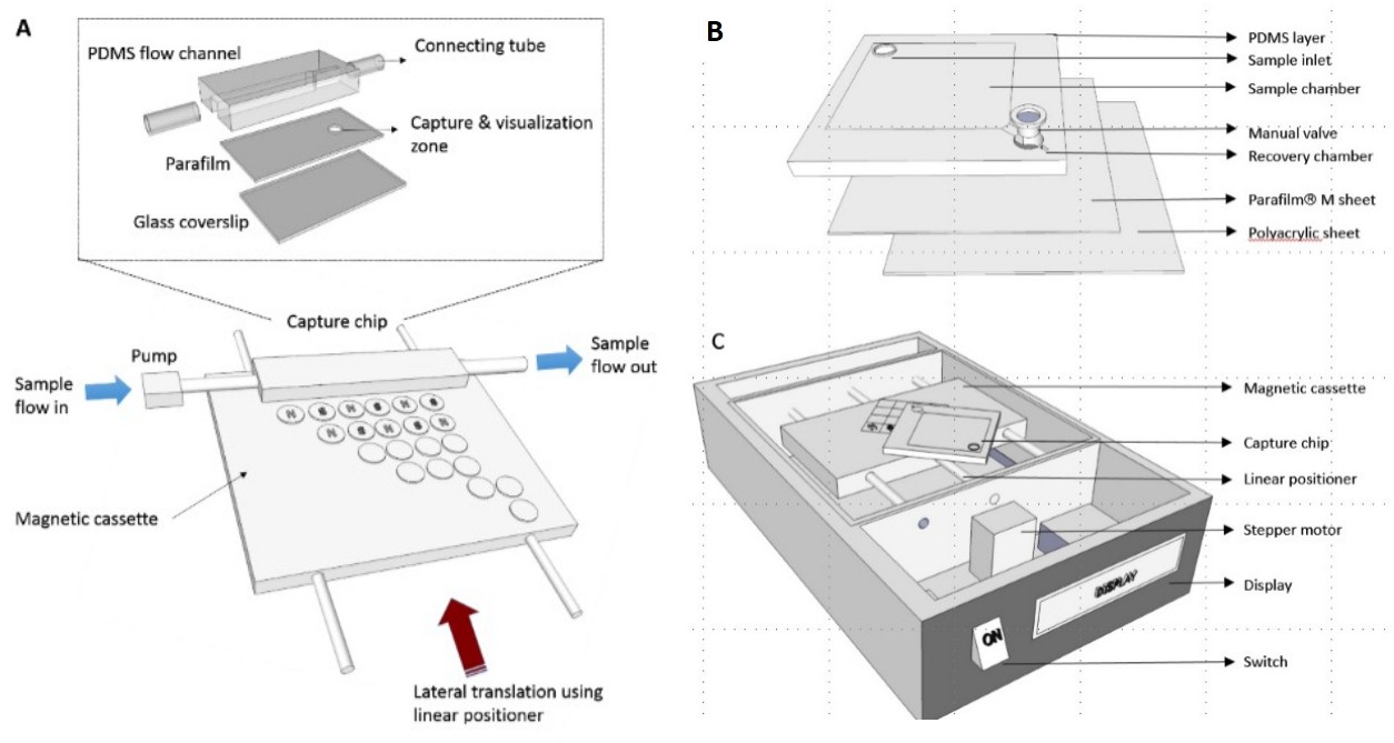
Detection of pathogens is particularly challenging when only a few cells per mL are present among a billion other blood cells. In collaboration with Dr. Ravi Elangovan’s and Dr. Vivek Perumal’s groups at IITD at We have developed two different formats of portable devices – SiMED and iMC2 which work on the principle of rapid immunomagnetic capture in a non-uniform field. This allows efficient enrichment of cells from 1 to 10 mL sample up to 100,000 times inside disposable millifluidic capture chips. The turnaround time for the devices is under 1 h using fluorescence microscopy/spectroscopy. Other downstream processes for detection that have been integrated with these devices are lateral flow assays, agglutination and impedance and allow different levels of sensitivity and processing times. The devices have been extensively validated on typhoid causing bacteria and are currently undergoing clinical trials. The devices are also being adapted for other diseases such as cholera and Tb.
(A) Spot immunomanetic enrichment device (SiMED), (B & C) Immunomagnetic cell capture (iMC2) system
- Singh S., Upadhyay M, Pandey V., Gupta S., Perumal V. and Elangovan R., ‘Spot Immunomagnetic Enrichment Device for Rapid Detection of Pathogens in Peripheral Blood‘, Advanced Materials Technologies, 1600101, 1 – 9 (2016)
- S. Singh, M. Upadhyaya, J. Sharma, S. Gupta, V. Perumal, and R. Elangovan, ‘A portable immunomagnetic cell capture system to accelerate culture diagnosis of bacterial infections‘ Analyst 141, 3358-3366 (2016)
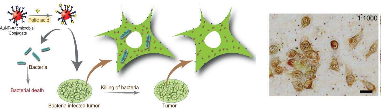
Project 3: Antibacterial approach via dual targeting in bacteria-infested cancer cells
Bacterial infections are showing ever increasing trends in multidrug resistance and have become a serious global threat to human society. Many pathogenic bacteria infect the patient through host cell-localization, evading the immune response and antimicrobial action of drugs, becoming more resistant over time. Cancerous tissues with hypoxic environment and chemo-attractants like purines provide a unique niche to bacteria and dogma holds that bacteria are causative agents of malignancy which is also supported by cellular studies at clinical levels. Chemotherapy can eradicate cancer cells but surviving bacteria can cause secondary infections in immunocompromised hosts leading to critical medical conditions. Thus, novel methodologies are urgently needed that allow facile modulation of antimicrobial action in a controlled manner to achieve higher avidity, bioavailability and internalization into mammalian cells. We are working closely with Dr. Neetu Singh’s group at IITD and have synthesized dual functionality peptide-gold nanobioconjugates that show enhanced antimicrobial activity (~ 40% higher than free drug) against Gram-negative bacteria intracellularized inside HeLa cells. We are currently working on more advanced strategies for target specific multivalent scaffolds which should further lower the antibiotic requirement as well as chances of drug resistance development.
- Singh R. and Gupta S., ‘Dual-functionality nanobioconjugates: A new tool for intracellular bacterial targeting in cancer?‘, Therapeutic Delivery, 9, 317-320 (2018)
- Singh R.*, Patil S.*, Singh N. and Gupta S., ‘Antimicrobial Activity of Polymyxin B Sulfate and Sushi Peptide Nanoparticle Bioconjugates on Gram-negative Bacteria’ Scientific Reports 7:5792, 1- 10 (2017) (*Equal contribution)
B. Bio/colloids in spatially-varying AC fields
The application of electric fields offers an effective and convenient way to dynamically measure, manipulate and control the assembly of colloidal systems in suspensions that is otherwise not readily achievable through techniques such as sedimentation, free volume restriction, capillary forces, convective evaporation and template-directed self-assembly. These structures can be harnessed for the fabrication of novel organic/inorganic mesoscopic functional materials and devices, potentially useful for sensing, catalysis, transport and medical purposes.
Project 1: Electrically-programmable assembly of colloids using dielectrophoresis (DEP)
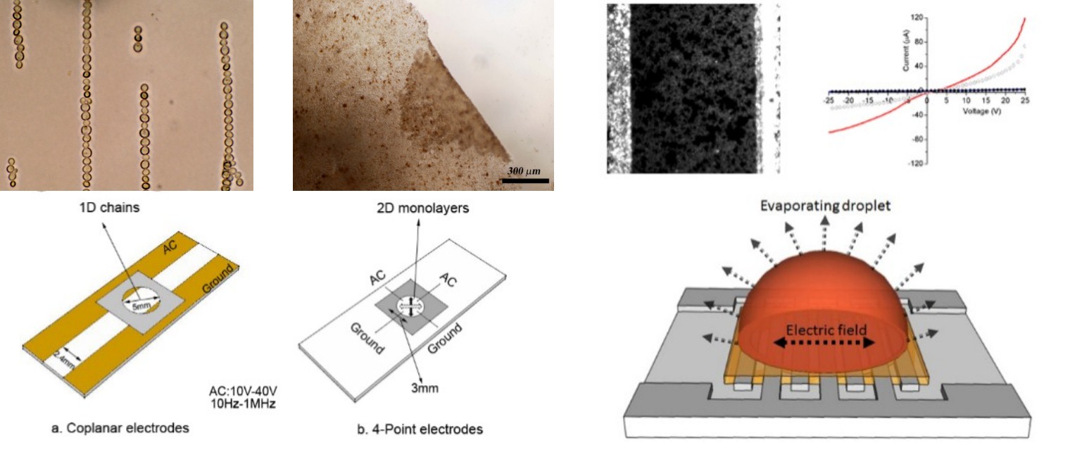
Our group has demonstrated the co-assembly of binary colloidal mixtures such as latex -latex or cells-latex into a variety of freely suspended (bio)active structures using DEP. The morphologies of the final structures can be controlled by tuning the voltage, frequency, pH, electrolyte and particle concentration. Using this approach, a variety of magnetically responsive biomaterials such as chains, arrays and membranes are prepared in which the assemblies are bound into permanent structures using different types of functionalized synthetic particles and ligands that attach to the cells through biospecific interactions. Simulations illustrate that the electric field draws the smaller sized particles between the larger ones which enables positioning of two particles types alternatingly.
Our group has also developed a simple, one-step strategy for the assembly of quantum dots (QDs) into photoconductive microwires using dielectrophoresis in AC electric fields on a chip. The fabrication is scalable as it does not require organic solvents, lengthy ligand-removal procedures, expensive microfabrication strategies (interdigitated electrodes with gap sizes as large as 100 microns may be used) or any sophisticated instrumentation. The optoelectronic structures can be readily used for photovoltaic applications.
Top row: DEP assembly of cell chains (left), cell-particle membranes (middle) and quantum dot microwires (right); Bottom row: Corresponding electrode geometry used for assembly.
- Maheshwari G., Mittal M., Sapra S. and Gupta S., ‘Electrically-Driven Assembly of CdTe Quantum Dots into Semiconducting Microwires’ J. Mater. Chem. C 3, 1645-1648 (2015)
- Jain S. and Gupta S., ‘Dielectrophoretic Co-Assembly of Binary Colloidal Mixtures in AC Electric Fields‘ Langmuir 29, 16105-16112 (2013)
- Gupta S., Alargova R.G., Kilpatrick P.K. and Velev O.D., ‘Directed Assembly of Live Cells into Functionalized Biomaterials using Dielectrophoresis on a Chip’ Langmuir 26, 3441-3452
- Gupta S., Alargova R.G., Kilpatrick P.K., and Velev O.D., ‘On-chip Electric Field Driven Assembly of Biocomposites from Live Cells and Functionalized Particles‘ Soft Matter 4, 726-730 (2008)
Project 2: Cellular microarrays
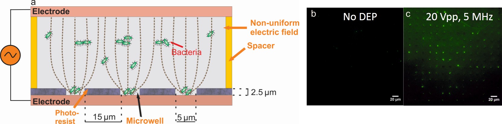
The principle of DEP has also been extended to capture of pathogenic bacteria into high-density miniaturized wells on a microfluidic chip that can be used for estimating the number of cells in binary mixtures or for antimicrobial susceptibility testing using impedance spectroscopy. Using a 34-mer antimicrobial peptide called sushi, the change in cell viability of S.typhi bacteria was detected with high sensitivity within an hour of antibiotic incubation which is currently not possible using disk diffusion or any other standard technique.
(a) Schematic of the experimental setup. Collection of S.typhi bacteria into microwells (b) before and (c) after applying AC electric field for 60 min.
- Goel M., Verma A. and Gupta S., ‘Electric-field driven capture and detection of live bacterial cells in microarrays‘, Biosensors and Bioelectronics, 11, 159 – 165 (2018)
Project 3: AC conductivity of colloidal suspensions
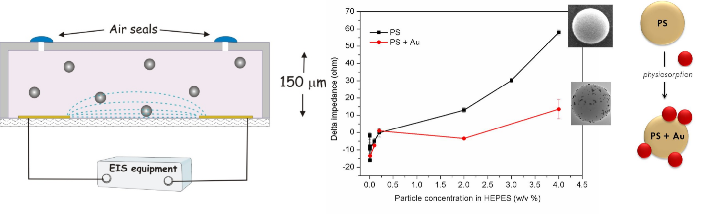
Impedance spectroscopy is explored as an efficient tool for probing the AC conductivity of colloidal suspensions and to design novel lab-on-chip biosensors. Our recent results demonstrate an exciting phenomenon wherein extremely dilute colloidal suspensions of different material types all show a characteristic non-monotonic conductive behavior that is uniquely dependent on the particle concentration. We are currently pursuing this work in greater detail in collaboration with Dr. Gaurav Goel’s group at IITD. We have also recently illustrated an immunosensor that can rapidly and sensitively detect infectious bacteria in 10 μL sample volumes. The bacteria are first tagged with gold nanoparticle bioconjugates via specific affinity antigen-antibody interactions and then subjected to high frequency AC electric fields applied via interdigitated microelectrodes. This allows massive signal amplification achieving a lower limit of detection (LOD) as 100 CFU/mL. These results are now being adapted for integration with iMC2 to accelerate the POC diagnosis of bacterial infections.
Left: Schematic of the microfluidic chip used for impedance measurements. Right: Non-monotonic impedance behavior for two different types of dielectric particle suspensions.
- Pal N., Sharma S. and Gupta S., ‘Sensitive and Rapid Detection of Pathogenic Bacteria in Small Volumes using Impedance Spectroscopy Technique’ Biosensors and Bioelectronics 77, 270-276 (2016)
- Gupta S., Kilpatrick P.K., Melvin E. and Velev O.D., ‘On-Chip Latex Agglutination Immunoassay Readout by Electrochemical Impedance Spectroscopy’ Lab on a Chip 12, 4279-4286 (2012)
C. Digital droplet microfluidics for fundamental and applied research
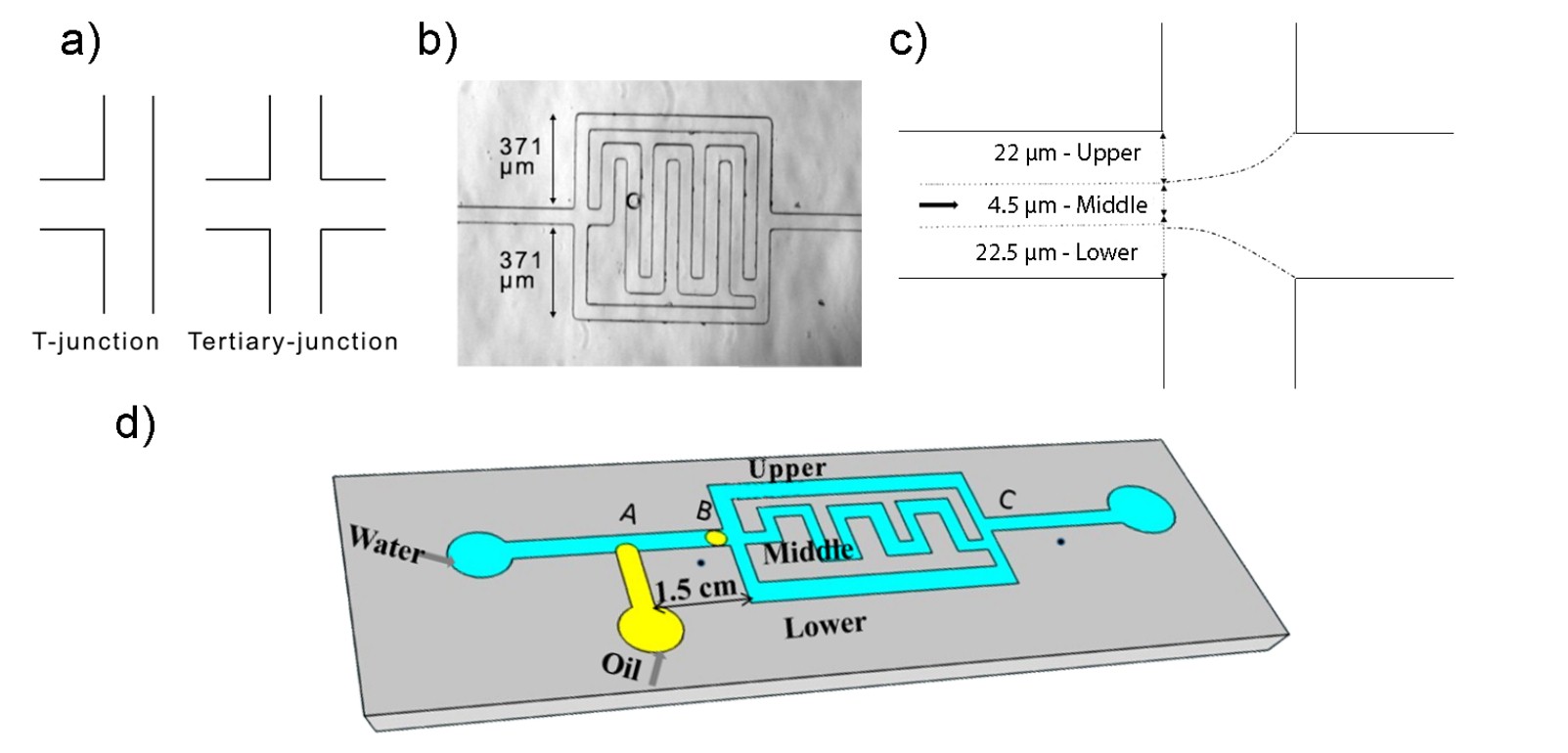
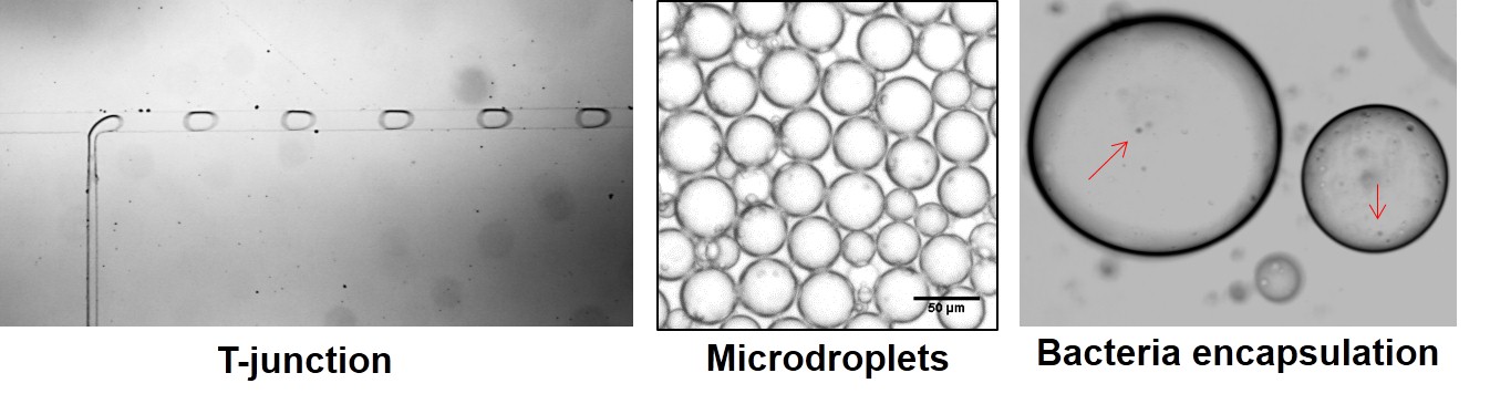
We work with droplet microfluidics in which picoliter volumes of oil-in-water (or water-in-oil) emulsions are generated in T- or flow focusing junctions and navigated through microfluidic channels for a variety of purposes ranging from flow characteristic studies at different operating conditions, microreactor synthesis to carry out biosensing work or for creating novel scaffolds for drug delivery and cellular applications.
Project 1: Droplet sorting inside tertiary-junction microchannels
We have investigated droplet navigation through microfluidic channels by studying the path selection by a single droplet inside a tertiary-junction microchannel using oil-in-water as a model system. A single droplet is generated at a T-junction inside a microfluidic chip and its flow behaviour as a function of droplet size, streamline position, viscosity, and Reynolds number (Re) of the continuous phase is studied downstream at a tertiary junction having perpendicular channels of uniform square cross-section and internal fluidic resistance proportional to their lengths. Both experimental and numerical simulations performed using the multicomponent lattice-Boltzmann method (LBM) suggest that at higher Re (~ 3), the flow is dominated by inertial forces resulting in the droplets choosing a path based on their center position in the flow streamline. At 10x lower Re, the streamline-assisted path selection become drag-force assisted above a critical droplet size. As the Re is further reduced (~ 0.03), or when the viscosity of the dispersed phase is increased, the critical droplet size for transition also decreases.
- Baig M.M., Jain S., Gupta S., Vignesh G., Singh V., Kondraju S. and Gupta S., ‘Engineering Droplet Navigation through Tertiary-Junction Microchannels’ Microfluidics and Nanofluidics, 20:165 (2016)
Project 2: Gel microreactors for drug delivery and biosensing
We are currently working on amyloid protein microgels and their “extrusion” using droplet microfluidics to form nanofibril microgel particles. These particles will be dielectrophoretically manipulated under spatially-varying variable AC fields and used as cell culture scaffolds and biogels for single-cell entrapment studies. We also envisage exploiting these microbeads as nanocontainers to encapsulate small molecules such as anti-bacterial and anti-cancer drugs for effective pharmacological action.
D. Triggered self-assembly of nanomaterials
Project 1: Agglutination assays for biomolecular detection
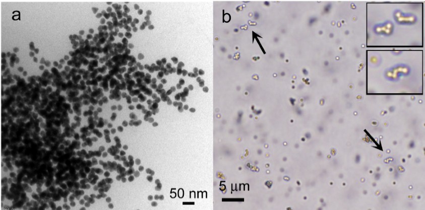
Biofunctionalized nanoparticles that can be triggered to aggregate on demand by inducing specific changes in their environment (pH, temperature, ions, biomolecules etc.) are extremely useful systems to study since their aggregation state can be readily monitored using standard optical techniques. Assembly of gold nanoparticles, for example, results in a characteristic red-to-blue shift in the visible spectrum. The ability to dynamically control the formation of these types of nanomaterials and to further make them tunable and/or switchable is valuable for both in vivo, in vitro sensing and drug delivery, and for engineering novel nanoparticle architectures. In the past, we have carried out experiments involving selective aggregation of peptide-functionalized gold nanoparticles in suspension for measuring the real-time activity and inhibitor screening of kinase enzymes. We are currently exploring the effects of nanomaterial assembly using specific biomolecular recognition of LPS-drug interactions.
(a) Kinase enzyme-actuated immunoaggregated network of gold nanoparticles. (b) Particle clusters formed via antibody-mediated latex-agglutination.
- Gupta S., Kilpatrick P.K., Melvin E. and Velev O.D., ‘On-Chip Latex Agglutination Immunoassay Readout by Electrochemical Impedance Spectroscopy’ Lab on a Chip 12, 4279-4286 (2012)
- Gupta S.*, Andresen H.A.* and Stevens M.M., ‘Kinetic Investigation of Bioresponsive Nanoparticle Assembly as a Function of Ligand Design’ Nanoscale 3, 383-386 (2011) *Equal contribution
- Gupta S., Andresen H.A. and Stevens M.M., ‘Single-Step Kinase Inhibitor Screening using a Peptide-Modified Gold Nanoparticle Platform’ Chemical Communications 47, 2249-2251 (2011)
- Gupta S., Andresen H.A., Ghadiali J.E. and Stevens M.M., ‘Kinase-Actuated Immunoaggregation of Peptide-Conjugated Gold Nanoparticles’ Small 6, 1509-1513 (2010)
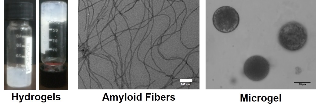
Project 2: Amyloid protein microgels
Amyloid proteins are formed as a result of protein aggregation (misfolding) wherein they undergo a conformational change from α-helix into β-sheets forming fibrillar structures. These amyloidic fibers are associated with several neurodegenerative diseases such as Alzheimer’s, Parkinson’s, Huntington and Type II diabetes, to name a few. This misfolding, although a major hazard from a clinical standpoint, can have remarkable implications for in vitro use if it is controlled properly under aqueous conditions. Under specific stimuli, amyloid fibril networks also interestingly form hydrogels that can be exploited in tissue engineering and regenerative medicine. Motivated by this study, our group is now working to explore synthetic amyloidogenic dipeptides and protein hydrogels with enhanced mechanical, electrical, and biological properties.